
Histology and Embryology
Research Focus
General Facts
Research
Selected Publications
Collaboration
Keywords: cellular electron microscopy, organelle ultrastructure, membrane trafficking, endosomal/lysosomal pathways, autophagy
Research (ÖSTAT Classification) : 106049, 106052, 301107, 301114, 301304
Research Focus
High-resolution microscopy with an emphasis on ultrastructural analysis of subcellular architecture in the context of intact tissues and cells from model organisms and patient samples.
General Facts
The Institute of Histology and Embryology at the Medical University of Innsbruck provides lecture series and practical courses in Histology and Microscopic Anatomy for undergraduate students in Human Medicine and Molecular Medicine. In addition, it offers special lectures and courses on submicroscopic morphology and on advanced cellular electron microscopy for MD and PhD students. The research of the cellular electron microscopy group focuses on ultrastructural aspects of intracellular membrane trafficking and signalling, in close collaboration with local and international research groups.
Research
The cellular electron microscopy group of M. W. Hess employs high-resolution microscopy for the ultrastructural analysis of subcellular architecture in the context of intact tissues and cells from model organisms and patient samples. The major focus of its cell biological studies is on intracellular membrane trafficking and signalling in health and disease (performed in collaboration with the groups of L. A. Huber and D. Teis – Division of Cell Biology; and T. Müller, A. Janecke and G. F. Vogel – Department of Paediatrics I) as well as the structural basis of bioadhesion (together with P. Ladurner and W. Salvenmoser – Department of Zoology, University of Innsbruck). Advanced cryo-based immunoelectron microscopy and electron tomographic 3D reconstruction are our methods of choice. We investigate (genome-edited) human and animal cell models (Perez Carion et al. 2018, Frontiers in molecular neuroscience 11:64; Yordanov et al. 2019, Traffic, 20:674), biopsy samples from patients (Vogel et al 2017, Traffic 18: 453) and eukaryotic model organisms such as mice, yeast (Adell et al. 2017, Elife 6:e31652), flatworms (Pjeta et al. 2019, Philosophical Transactions of the Royal Society B 374:20190194) and Hydra (Rodrigues et al. 2016, BMC Zoology 1:3). Membrane trafficking is studied with special emphasis on the biogenesis and maturation of endocytic compartments, cargo biosynthesis and recycling as well as autophagy. Among other things, we are also interested in the relationships between cargo biosynthesis and trafficking, cytoskeletal architecture and the maintenance of cellular polarity. All these processes are severely disturbed in the case of microvillus inclusion disease (MVID), a rare, fatal, congenital, intestinal disease that affects infants soon after birth. The clinical appearance presents severe watery, non-inflammatory diarrhoea, nutrient malabsorption and metabolic acidosis. In general, MVID patients depend on total parenteral nutrition. Small bowel transplantation is the only curative therapy but many patients die within the first few years of life. In previous years, our multidisciplinary cell biological-clinical team provided mechanistic insights into the pathophysiology of MVID. We identified mutations in the motor protein myosin 5b, in the apical membrane protein syntaxin 3 or in its interaction partner syntaxin-binding protein 2 as causative of this disease. These mutations disrupt selective apical cargo trafficking and exocytosis in epithelial absorptive cells of the small intestine, leading to mislocalisation of pivotal brush border ion transporters relevant for physiological enterocyte function (T Müller et al. 2008, Nature genetics 40:1163; Wiegerinck et al. 2014, Gastroenterology 147:65; Vogel et al. 2017, JCI-Insight 2(14): e94564; Vogel et al 2017, Traffic 18: 453). Finally, we perform methodological research for the development of new preparation and imaging techniques, for the precise 3-dimensional localisation of macromolecules in the context of natively cryo-immobilised cells (Hess et al. 2018, Traffic 19:639).
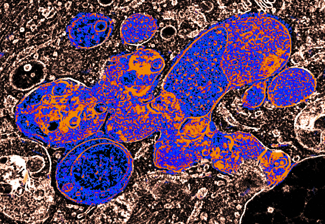
Fig. 1: Pleiomorphic, large endo-lysosomal compartments, as seen in cryofixed HT1080 human fibrosarcoma cell cultures. Pseudo-colour electron microscopy image.
Selected Publications
- Yordanov, Teodor E.; Hipolito, Victoria E. B.; Liebscher, Gudrun; Vogel, Georg F.; Stasyk, Taras; Herrmann, Caroline; Geley, Stephan; Teis, David; Botelho, Roberto J.; Hess, Michael W.; Huber, Lukas A.: Biogenesis of lysosome-related organelles complex-1 (BORC) regulates late endosomal/lysosomal size through PIKfyve-dependent phosphatidylinositol-3,5-bisphosphate. TRAFFIC. 2019; 20(9); 674-696. doi: 10.1111/tra.12679
- De Araujo, Mariana E.G.;Liebscher, Gudrun; Hess, Michael W.; Huber, Lukas A.: Lysosomal size matters. TRAFFIC. 2020; 21(1); 60-75.doi: 10.1111/tra.12714
- Schmidt, Oliver; Weyer, Yannick; Sprenger, Simon; Widerin, Michael A.; Eising, Sebastian; Baumann, Verena; Angelova, Mihaela; Loewith, Robbie; Stefan, Christopher J.; Hess, Michael W.; Fröhlich, Florian; Teis, David: TOR complex 2 (TORC2) signaling and the ESCRT machinery cooperate in the protection of plasma membrane integrity in yeast. THE JOURNAL OF BIOLOGICAL CHEMISTRY. 2020; 295(34); 12028. doi: 10.1074/jbc.RA120.013222
- Wunderer, Julia; Lengerer, Birgit; Pjeta, Robert; Bertemes, Philip; Kremser, Leopold; Lindner, Herbert; Ederth, Thomas; Hess, Michael W.; Stock, David; Salvenmoser, Willi; Ladurner, Peter: A mechanism for temporary bioadhesion. PROCEEDINGS OF THE NATIONAL ACADEMY OF SCIENCES OF THE UNITED STATES OF AMERICA. 2019; 116(10); 4297-4306. doi: 10.1073/pnas.1814230116
- Pjeta, Robert; Wunderer, Julia; Bertemes, Philip; Hofer, Teresa; Salvenmoser, Willi; Lengerer, Birgit; Coassin, Stefan; Erhart, Gertraud; Beisel, Christian; Sobral, Daniel; Kremser, Leopold; Lindner, Herbert; Curini-Galletti, Marco; Stelzer, Claus-Peter; Hess, Michael W.; Ladurner, Peter: Temporary adhesion of the proseriate flatworm Minona ileanae. PHILOSOPHICAL TRANSACTIONS OF THE ROYAL SOCIETY B-BIOLOGICAL SCIENCES. 2019; 374(1784); 20190194. doi: 10.1098/rstb.2019.0194]
Collaborations
- Peter Ladurner, Bert Hobmayer (Department of Zoology, University of Innsbruck, Austria)
- James R. Goldenring (Section of Surgical Sciences, Epithelial Biology Center, and Department of Cell & Developmental Biology, Vanderbilt University School of Medicine, Nashville, TN, USA)
 Univ.-Prof. Dr.med.univ. Lars Klimaschewski
Director
Contact:
Müllerstrasse 59
6020 Innsbruck
Austria
Email: lars.klimaschewski@i-med.ac.at
Phone: +43 512 9003 71160
https://www.i-med.ac.at/ahe/histologie-embryologie/