
Neuroradiology
Research Focus
General Facts
Research
Selected Publications
Selected Funding, Collaboration
Devices & Services
Keywords: High-field MRI, MR-spectroscopy, fMRI, DTI, multimodal imaging, quantitative MR-imaging, artificial intelligence, dual-energy-CT, interventional neuroradiology, dose management
Research (ÖSTAT Classification) : 301111, 301401, 302010, 302043, 302044, 302070, 302071, 302079
Research Focus
To a large extent, neuroradiological research is connected with and driven by its clinical partners (Neuro-Focus at MUI) as well as other departments (clinical and theoretical) at MUI, MCI, UMIT and LFU. Projects generated within Neuroradiology focus mostly on technical developments (dose reduction / dual-energy CT, MRI sequence and post-processing developments, fMRI/VBM, multi-nuclear MRI), some in cooperation with the Department of Radiology. Furthermore, Neuroradiology conducts clinical studies, e.g. into neurovascular disease (Fig. 1).

Fig. 1: Endovascular treatment of cerebral aneurysms is becoming increasingly important in clinical routine. The first row shows a recent development in stents, which is evaluated here in clinical studies. The second row shows an example of endovascular stroke treatment with thrombectomy, using one of the recently developed stent retrievers (arrows). Neuroradiology (in cooperation with Neurology) is part of a large multi-centre study here
General Facts
Structure of the Research Unit, Aims and Clinical Routine
Together with the Department of Radiology, Neuroradiology is involved in radiographer and physician training programmes as well as research and routine clinical activities. The two departments have a long-standing PhD programme (Image-Guided Diagnosis & Therapy) and a clinical PhD programme (Clinical Imaging Science).
Neuroradiology has a large clinical workload, which includes all diagnostic and interventional neurology cases of paediatric and adult patients, resulting in many clinically related research projects in imaging and intervention (including interventional EU studies). The senior physicians and one Assoz. Professor lead the younger colleagues and the PhD students in all aspects of research and education, supported by the scientists of the Core Facility for Neuroimaging Research (CF-NIR) including MR physics, advanced mathematics and artificial intelligence. This leads to new insights in scanning, image analysis and follow-up predictions.
The experimental research relates mainly to the CF-NIR but also to many national and international cooperative partners. There is long-standing expertise in fMRI, ultra-high field MRI (7T in cooperation with the high-field MRI excellence centre at MUW and with the imaging unit of DKFZ Heidelberg), multimodal, multi-nuclear and quantitative imaging. Imaging of cerebral processing related to cognitive and emotional stimuli with respect to possible gender differences relates partly to psychiatric research and partly to projects in critical emotional situations in cardiology.
Research
This section lists only those research projects that are led mainly by Neuroradiology.
Dual-Energy CT in Stroke Patients
Assoz.-Prof. Grams, Dr. Janjic, Dr. Mangesius, Dr. Pereverzyev in cooperation with Prof. Kiechl, Prof. Knoflach and Prof. Haltmeier (LFU)
The detection of acute cerebral infarction is crucial in order to decide on further treatment. By comparison with conventional CT, early infarctions can be identified better with dual-energy CT (Fig. 2). A recent research project aims to predict infarction development prior to its visualisation, by using radiomics features in dual-energy CT datasets.
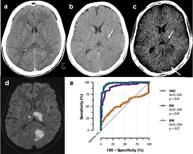
Fig. 2: Optimised visualisation of early cerebral infarction with dual-energy computed tomography. With a conventional brain window (BM, a) series, no infarction is visible. With a dual-energy “virtual non-contrast” (VNC, b) reconstruction, frequently used to detect cerebral haemorrhage, a left basal ganglia infarction can be assumed (arrow). With a dedicated “oedema map” (EM, c) reconstruction, a left basal ganglia and occipital infarction can be detected (arrows), which corresponds to the final infarctions, seen with diffusion-weighted MRI (d). With EM, early cerebral infarctions can be visualised with a high level of sensitivity and specificity (e).
Aneurysm Wall Enhancement in Cerebral Aneurysms
Assoz.-Prof. Dr. Grams, Dr. Ladenhauf and Prof. Gizewski in cooperation with PD Dr. Helbok
This project involving patients with treated or untreated cerebral aneurysms is to investigate aneurysm wall enhancement. The presence, frequency and severity of enhancement under different circumstances and correlation with aneurysm reperfusion will be investigated
MRI and MRS Parameters in the Cerebral Development of Preterm Infants
Dr. Djurdjevic, Prof. Gizewski and Dr. Pereverzyev in cooperation with Prof. Kiechl-Kohlendorfer and Prof. Buchheim (LFU)
In this prospective study into multimodal MRI in very preterm neonates (< 32 gestational weeks), we employed feed-forward neural networks (fNNs), a segment of artificial intelligence. We showed that fNNs might be a useful predictive tool for cognitive and motor outcome prediction in very preterm neonates when using cerebral 1H-MRS and DTI biomarkers (Fig. 3). Moreover, by combining these sophisticated methods, the predictive value of MRI can be significantly increased. Further applications are now being tested in a larger cohort.
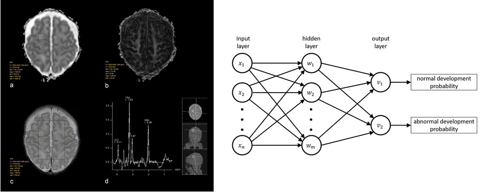
Fig. 3: An example of region-of-interest-based evaluation of ADC (a) and FA (b) in frontal white matter on the right side with a corresponding voxel in 2D CSI 1H-MR spectroscopy (c, d).
A network diagram on the right, representing the neural networks employed in the MRS and DWI data of preterm children.
Each unit of the input and hidden layer connects to each unit of the subsequent layer. Through these connections, every unit of the hidden and output layer is a linear combination of all units of the preceding layer followed by a non-linear transfer function. The input units xj correspond to the variables used for class prediction (metabolite ratios and DTI characteristics).
31P MRS in Cerebral Gliomas and Metastases
Assoz.-Prof. Grams; Prof. Gizewski, Dr. Walchhofer and Dr. Steiger in cooperation with Prof. Thomé, Dr. Kerschbaumer, Doz. Freyschlag, Prof. Stockhammer, Doz. Nowosielski and Prof. Nevinny-Stickel
By using MR spectroscopy of phosphorus compounds (31P MRS), it is possible to detect metabolites of energy metabolism and of membrane turnover. 31P MRS is used in patients with cerebral gliomas and metastases, to investigate tumour heterogeneity and the effects of therapy not only on the tumorous area but also on the healthy brain hemisphere. The resulting data will be correlated with results obtained from established methods, such as 1H MRS, MR perfusion, DWI, and clinical, histological and PET parameters.
31P MRS in Stroke Patients
Assoz.-Prof. Grams, Dr. Walchhofer, Dr. Rietzler and Dr. Steiger in cooperation with Prof. Kiechl and Prof. Knoflach
31P MRS is used in patients with acute, subacute and chronic ischemic stroke, to gain further insights into the energy metabolism and reorganisation mechanisms of infarcted brain and surrounding areas during the acute stage and to monitor subacute and chronic changes.
Contribution of Myelin to the Diamagnetic Susceptibility of Normal and Multiple Sclerosis (MS) Brain
DI Dr. Birkl (Erwin-Schrödinger Fellow) in cooperation with Prof. Haybäck
Although a wide spectrum of MRI techniques is available to assess the density and integrity of myelin, the detailed contribution of its different constituents in relation to magnetic susceptibility is still understood only partially. A novel technique called quantitative susceptibility mapping (QSM) allows assessment of the magnetic susceptibility of biological tissue using MRI. The strongest contributors to magnetic susceptibility of the human brain are iron and myelin. The main aim of this project is to uncover the influence of myelin (myelin water imaging – MWI) and its different components on the magnetic susceptibility of healthy and MS brain tissue.
Imaging Markers for Tissue Damage and Disease Progression in MS
DI Dr. Birkl and Prof. Rauscher (University of British Columbia, Canada)
There is still a lack of advanced MRI tools for early MS diagnosis, monitoring of disease progression and quantification of tissue repair from MS drugs currently under development. Our broad aim is to quantify tissue damage in MS by means of QSM and MRI signal decay (R2*). The aim of this project is to establish white matter fibre orientation-dependent R2* as an imaging marker for disease progression in MS. For this approach, we calculate R2* as a function of white matter fibre orientation. By matching a computational model of iron and myelin to experimental data, we are able to determine average total content of white matter iron and myelin.
Towards Quantitative Neuroimaging Biomarkers for Friedreich’s ataxia at 7 Tesla
Prof. Gizewski and Dr. Mangesius in cooperation with the German Cancer Research Centre, Heidelberg (Dr. Straub and Prof. Ladd) and PD Dr. Bösch (Neurology Innsbruck)
Modern imaging processes and new imaging techniques with ultra-high field 7T MRI are being investigated. FRDA leads to degenerative cerebral processes. So far, no effective treatment has been found. It is therefore important to aid the development of medication with imaging biomarkers that reflect disease status and progress. MRI data for QSM, R1, R2* and DWI have been acquired at 7T and have facilitated the identification of statistically significant differences between FRDA patients and controls in five out of twelve brain structures (Fig. 4) investigated, which also correlated positively with disease characteristics. Future studies will include the application of these markers in routinely applied 3T field strengths and the investigation of asymptomatic mutation carriers.
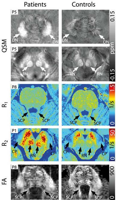
Fig. 4: Representative axial slices of susceptibility maps (QSM), colour-coded diffusion of fractional anisotropy maps (DW-cFA), T2 and T1 maps depicting the superior cerebellar peduncle (black arrow), nucleus ruber and sustantia nigra (white arrows) in one patient (first column) and one healthy control (second column).
Cerebral Processing of Food Stimuli in Young Anorexic Patients with Respect to Personality Disorders and Gender
Prof. Gizewski and Dr. Steiger in cooperation with Prof. Sevecke and Dr. Gander
Some earlier studies revealed alterations in cerebral processing in adult anorexic patients. However, since these were based on long-standing disease, the results were able to provide no clear answers on how these functional and structural differences developed in contrast with healthy volunteers. We have therefore established the application of these stimuli to young patients and will correlate the measured brain parameters with psychosocial data.
Effective Dose Assessment for Fluoroscopically Guided Interventional Procedures: Frequency of Dose Above 100 mSv in a European Centre
Prof. Gizewski, Prof. Jaschke, Dr. Verius, Dr. Eder and Dr. Torbica in cooperation with Prof. Rehani (Radiology Boston, USA)
In interventional radiology, the use of fluoroscopically guided interventional (FGI) procedures is increasing. Dose management is crucial, as indications vary over all body areas and there is a broad range of intervention techniques. The purpose of this study is to investigate the distribution of patients who received a substantial cumulative effective dose (CED) over different FGI procedures, using the semi-automated dose-tracking system developed in-house.
Effect of Short-Term Meditation Training on Brain Metabolism, Function and Volume
Prof. Gizewski, Dr. Steiger, Dr. Lenhart, Dr. Mangesius, Dr. Siedentopf, Dr. Pereverzyev and Dr. Galijasevic in cooperation with Prof. Singewald (LFU)
Meditation is increasingly attracting interest among neuroimaging researchers on the basis of its relevance as a cognitive enhancement technique. This longitudinal study applies a distinct and standardised meditative technique with a group of volunteers in a short-term training programme, in order to analyse brain metabolic, functional and structural changes. Initial results indicate that meditation may have considerable effects on brain energy state with different local energy management in specific brain regions and may alter cerebral grey and white matter mainly in the insula, caudate nucleus, frontal cortices, parietotemporal regions, right medial prefrontal cortex, basal ganglia and parahippocampal gyrus (Fig. 5).
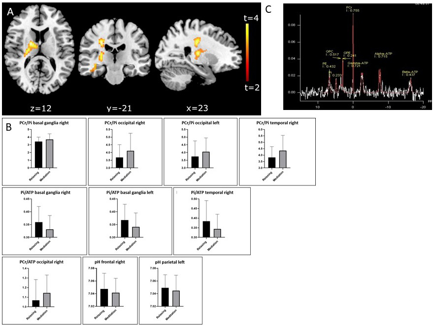
Fig. 5: A: Statistical parametric mapping rendered on a normalised MRI scan showing increases in areas of significant fractional anisotropy in individuals after focused attention meditation training compared with the time point before training (FEW-corrected at P < 0.05 where height threshold P < 0.01).
B: Bar graphs with mean values and standard deviations of all significant differences in 31P MR spectroscopy (p < 0.00125) after meditation training.
B: Typical 31P MR spectrometry in a normal brain
Selected Publications
- Straub S; Mangesius S; Emmerich J; Indelicato E; Nachbauer W; Degenhardt KS; Ladd ME; Boesch S; Gizewski ER: Towards quantitative neuroimaging biomarkers for Friedreich's ataxia at 7 Tesla: susceptibility mapping, diffusion imaging, R2 and R1 relaxometry. J Neurosci Res, 2020 Jul 30; 98(11): 2219-31.
- Janjic T; Pereverzyev S Jr; Hammerl M; Neubauer V; Lerchner H; Wallner V; Steiger R; Kiechl-Kohlendorfer U; Zimmermann M; Buchheim A; Grams AE; Gizewski ER: Feed-forward neural networks using cerebral MR spectroscopy and DTI might predict neurodevelopmental outcome in preterm neonates. Eur Radiol. 2020 Dec; 30(12): 6441-6451.
- Diechtl W, Tuovinen N, Barbieri F, Adukauskaite A, Senoner T, Rubatscher A, Hintringer F, Siedentopf C, Bauer A, Gizewski ER, Steiger R: Functional neuroimaging in the acute phase of Takotsubo syndrome: volumetric and functional changes oft he right insular cortex. Clin Res Cardiol. 2020 Sep;109(9): 1107-1113.
- Nigro S, Antoinini A, Vaillancourd DE, Seppi K, Ceravolo R, Strafella AP, Augimeri A, Quattrone A, Morelli M, Weis L, Fiorenzato E, Biundo R, Burciu RG, Krismer F, McFarland NR, Mueller C, Gizewski ER, Cosottini M, Del Prete E, Mazzucchi S, Quattrone A: Automated MRI Classification in Progressive Supranuclear Palsy: A Large International Cohort Study. Mov. Disord. 2020 Jun;35(6):976-983.
- Sporns PB, Sträter R, Minnerup J, Wiendl H, Hanning U, Chapot R, Henkes H, Henkes E, Grams A, Dorn F, Nikoubashman O, Wiesmann M, Bier G, Weber A, Broocks G, Fiehler J, Brehm A, Psychogios M, Kaiser D, Yilmaz U, Morotti A, Marik W, Nolz R, Jensen-Kondering U, Schmitz B, Schob S, Beuing O, Götz F, Trenkler J, Turowski B, Möhlenbruch M, Wendl C, Schramm P, Musolino P, Lee S, Schlamann M, Radbruch A, Rübsamen N, Karch A, Heindel W, Wildgruber M, Kemmling A: Feasibility, Safety, and Outcome of Endovascular Recanalization in Childhood Stroke: The Save ChildS Study. JAMA Neurol. 2020 Jan 1; 77(1): 25-34
Selection of Funding
- National cooperation partner FWF: KLI 543-B27 (Prof. Trinka, Salzburg: “Emotionserkennung und soziale Kognition bei JME Patienten” (Emotion recognition and social cognition in JME patients) .
- EU H2020-SC1-2016-2017, TENSION Study (Efficacy and safety of thrombectomy in stroke with extended lesion and extended time window: a randomised controlled trial) (Gizewski: Austrian PI)
- FFG COMET programme: Gizewski: PI Subproject “Imaging biomarkers for vascular disease and vascular ageing”, K1 Centre Tirol: VASCage-C (Centre for the Promotion of Vascular Health in the Ageing Community)
- Mangesius and Schiefeneder anniversary fund: “Automatisierte MR-Planimetrie-Messungen zur Diagnose und Prognose von Multipler Sklerose” (Automated MR planimetry measurements for multiple sclerosis diagnosis and prognosis)
- ÖAW (Mangesius, Gruber, Posch and Irschara in cooperation with Radiology and the Department of Language and Literature, LFU): Retrospective Intersectional Corpus-Linguistic Analysis of Radiology Reports from Innsbruck, Medical University, MedCorpInn
- FWF P project: Pereverzyev: “Regularisation techniques in learning with big data”
Collaborations
- Austria: NEUROIMAGE WING (BMWFW grand: pooled MRI data collection and analysis Med. Universities Innsbruck, Graz, Vienna (Neurology and Neuroradiology, 7T MRI)
- Innsbruck: Prof. Baumgarten, Biomedical Informatics and Mechatronics (UMIT)
- Prof. Markus Haltmeier, Applied Mathematics (LFU), Prof. Matthias Harders, Interactive Graphics and Simulation (LFU)
- Prof. Anna Buchheim and Team, Clinical Psychology (LFU)
- Germany: University Hospital Essen (Medical Psychology & Behavioural Immunobiology, Radiology), Erwin L. Hahn Institute Essen (Prof. Harald Quick)
- DKFZ Heidelberg / MR Imaging (Prof. Mark Ladd)
- Dresden Technical University (Prof. Jennifer Linn, Neuroradiology), Hamburg University (Prof. Jens Fiehler, Neuroradiology)
- Switzerland: Hirslanden Clinic Zürich, Prof. Isabel Wanke, Prof. Daniel Rüfenacht, Neuroradiology
- Canada: University of British Columbia (Biomedical Imaging)
- Siemens Healthineers (Erlangen, Germany), Kinepict Health Ltd. (Budapest, Hungary)
Devices & Services
Neuroimaging Research Core Facility
The main modality of this CF is the BMWF-funded 3-Tesla MRI system, which has established a core facility for MR-based neuroimaging research at MUI. The 3T MRI started out exclusively for research use in 2012. The CF NIR is centrally administered by the Head of the Department of Neuroradiology, who leads an interdisciplinary steering board. The technical equipment is supported by one physicist, one mathematician and 2 assistant radiographers. The Neuroradiology team supports all associated scientists on technical and post-processing questions. Furthermore, the core facility develops and introduces new MR sequences and technical equipment (currently sodium imaging of the brain). Above all, the Neuroimaging platform offers opportunities to bring different groups together and to transfer knowledge as well as providing a setting for communication and cooperation (e.g. within the Neuroimage WING, a grant – higher education structural funding – to fund an imaging platform at the Medical Universities of Innsbruck, Graz and Vienna).
 Univ.-Prof.in Dr.in Elke R. Gizewski MHBA
Director
Contact:
Anichstraße 35
6020 Innsbruck
Austria
Email: neuroradiologie@i-med.ac.at
Phone: +43 512 504 27095
Fax: +43 512 504 27096
http://radiologie.tirol-kliniken.at