
Orthodontics
Research Focus
General Facts
Research
Selected Publications
Selected Funding, Collaboration
Devices & Services
Keywords: Artefacts of brackets, DECT, surface-coated materials, bioactive, aligner treatment, maxillary expansion, orthodontic tooth movement, pro-inflammatory substances
Research (ÖSTAT Classification) : 302033, 302034, 302044, 302071, 301108, 211904
Research Focus
- Reduction of artefacts of brackets using dual-energy CT (DECT) by creating monochromatic reconstructions (MC) in the tooth and jaw area.
- Evaluation of osseointegration on loaded, strontium-functionalised orthodontic mini screws (Ti-Sr-O).
- Precision of aligner (Invisalign®) treatment with the current material (SmartTrack®) in achieving expansion or contraction of the maxilla and occlusal contacts.
- Systemic use of certain pro-inflammatory substances to accelerate orthodontic treatment.
General Facts
The University Hospital for Orthodontics is part of the Department of Dental and Oral Medicine and Cranio-Maxillofacial and Oral Surgery. There is no extra laboratory staff.
The major aim of our basic and clinical research activities in the field of orthodontics is to increase knowledge in the respective research areas and to optimise patient treatment.
The current research topics of the University Hospital for Orthodontics focus on strontium-functionalised orthodontic mini screws, the reduction of artefacts of brackets using dual-energy CT (DECT), and investigation of the precision of aligner treatment. Further clinical research focuses on the role of pro-inflammatory substances in accelerating orthodontic treatment.
We collaborate closely with a number of clinical and scientific units at the Medical University of Innsbruck as well as international research partners. Currently, there are strong collaboration activities on the Innsbruck campus with the Institute of Clinical and Functional Anatomy, the University Hospital for Cranio-Maxillofacial and Oral Surgery, the University Hospital for Radiology and the Institute of Pathology, Neuropathology and Molecular Pathology. For outside collaborations and core facilities, see below.
Research
Potential of Artefact Reduction of Brackets using Dual-Energy CT
Manuel Kasslatter
Because of the high atomic numbers and high density of the materials used in orthodontics, maxillofacial surgery and tooth preservation, beam hardening artefacts represent a major problem in computed tomography (CT) imaging of the teeth and jaw.
The aim of this work is to examine the aforementioned artefacts and to reduce them by creating monochromatic reconstructions (MC) in the tooth and jaw area.
Dual-energy computed tomography (DECT) is performed on ten anatomical specimens, i.e. corpse heads, with and without orthodontic equipment comprising a vacuum-formed template with incorporated brackets and an archwire (Fig. 1). All the dental arches exhibit metal restorations to a greater or lesser extent, which themselves have also caused artefacts. MC from 80 to 160 kV are then generated. The artefacts are assessed qualitatively and quantitatively using established methods. In anatomical structures such as the floor of the mouth and the soft palate, which are not in the same axial plane as objects with a high atomic number that produce artefacts, an increase in Hounsfield Units (HU) can be observed as the kV number of the MC increases. In structures on the same level as objects that produce artefacts, a decrease in HU can be observed as the kV number of MC increases.
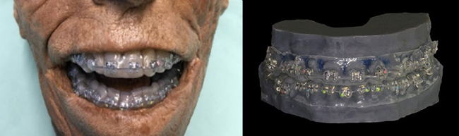
Fig. 1: Corpse head and SL 3D-printed model with orthodontic equipment comprising a vacuum-formed template with incorporated brackets and an archwire.
This study shows that MC artefact reduction is also possible in the jaw area. In all studies, no significantly increased artefacts are found in the setting with orthodontic appliances, in comparison to the setting without orthodontic appliances. Other than artefacts as a result of metal restorations in the teeth, no significant differences can be found between setting with and without orthodontic appliances in any of the ten ROIs groups examined.
Osseointegration of Strontium-Coated Orthodontic Mini Screws Under Load
Natalie Schenz
Mini screws have become an important tool in orthodontic practice. The objective of this study is to evaluate osseointegration via bone-to-implant contact and peri-implant bone formation on loaded, strontium-functionalised, orthodontic mini screws (Ti-Sr-O).
Mini screws (titanium Ti and strontium-coated Ti-Sr-O) are inserted monocortically into either tibia of 10 male Wistar rats and a force of 25 cN is immediately applied through a coil spring. After a two-week healing period, specimens are harvested and analysed via histomorphometry. Bone-to-implant contact (BIC%) and peri-implant bone formation (BF%) in defined regions of interest (ROI-I; ROI-II) are measured on both sides of the implant (Fig. 2).
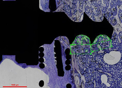
Fig. 2: Histological evaluation of bone implant contact (BIC) and peri-implant bone formation (BF) caudally at the mini screw.
Ti-Sr-O-functionalised implants show significantly more bone formation in ROI-I than in the control group with Ti mini screws on the pulling side. With respect to the pushing side, greater bone formation is observed but with no statistical significance. Moreover, BIC shows no statistically significant differences between the groups.
The study presented shows that, even under loading conditions, strontium-functionalised mini screws have greater bone formation than regular titanium mini screws und could therefore serve as a modification for temporary anchorage devices in orthodontics. Current research is focusing on the osseointegration of strontium-coated, orthodontic mini screws under load after longer healing periods of four and six weeks. In addition, osseointegration will be examined by means of a synchrotron scan.
Maxillary Expansion or Contraction and Occlusal Contact Adjustment: Effectiveness of Current Aligner Treatment
Ulrike Riede
The aim of this study is to evaluate the precision of aligner (Invisalign®) treatment with the current material (SmartTrack®) in achieving expansion or contraction of the maxilla and occlusal contacts, as simulated in the proprietary planning software (ClinCheck®, CC).
Thirty patients thus treated are evaluated retrospectively. Four maxillary models are analysed per patient: a pre-treatment model, a scan-based CC model, a post-treatment clinical model, and a CC model reflecting the treatment outcome as initially simulated. Thirteen transverse parameters are measured separately on each model by two investigators. Occlusal contacts are also analysed (Fig. 3).
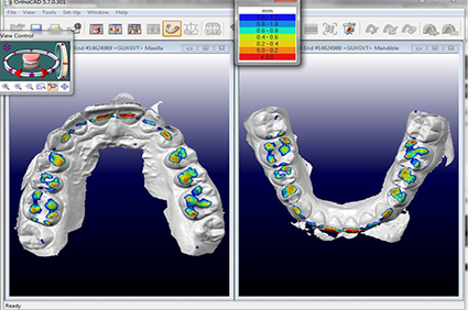
Fig.3: OrthoCAD: digital models show occlusal contact adjustment.
The measuring method is validated by both investigators arriving at similar results for the effectiveness with which the simulated treatment goals has been clinically achieved. Significant differences are observed in transfer precision from the casts to the planning software and between the simulated and clinical outcomes. Intense occlusal contacts in the simulations materialise less frequently (≈ 2%) than ideal contacts (≈ 60%) in the clinical outcomes.
The effectiveness of achievement of the simulated transverse goals is 45% and generally not found to be better with SmartTrack® than with the previously used Ex30® material. Out of 100 simulated occlusal contacts, 40 will never materialise and the achievement of around 60 will adequately ensure a clinically favourable contact pattern.
With the caveat that any overcorrection will reduce precision to some extent, it seems perfectly possible to make deliberate use of overcorrection in current aligner therapies for transverse maxillary expansion or contraction.
Effects of Different Oral Therapeutics on Orthodontic Tooth Movement
Lisa Schieffer
The aim of every orthodontic treatment is to move teeth efficiently and gently at the same time. The speed of orthodontic tooth movement is multifactorial. On the one hand, it is dependent on the physiological conditions in the periodontal tissue, which in turn are subject to the control of molecular mechanisms that regulate cellular behaviour in the alveolar bone and the periodontal ligament. On the other hand, it is dependent on the externally applied orthodontic forces, subject to the equipment used (Fig. 4).
Biological research at tissue, cell and molecular level has revealed the possibility of accelerating orthodontic tooth movement by adding certain mediators. This is still the subject of numerous scientific studies with the aim of shortening the duration of orthodontic treatment and thus reducing possible side effects.
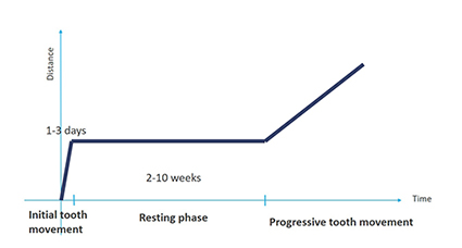
Fig. 4: Phases of orthodontic tooth movement. Phase I: deviation of tooth and compression of PDL; Phase II: hyalinization; Phase III: progressive tooth movement.
It has been proven that proinflammatory agents in particular have a positive effect on the differentiation of osteoclasts, which accelerates tooth movement. In the future, local or systemic use of certain pro-inflammatory substances could play an important role in accelerating orthodontic treatment. Further studies will be needed to clarify the role of metabolites in achieving maximum tooth movement whilst minimising root damage during orthodontic tooth movement.
Selected Publications
- Schenz N, Schwarz V, Hörmann R, Crismani AG: Impression material accuracy for palatal orthodontic miniscrews. JOURNAL OF OROFACIAL ORTHOPEDICS: 2020; 81: S. 427-439
- Kasslatter M, Glodny B, Grams A, Crismani A, Schieffer L: Pilot Study: Comparison of Artefacts of Brackets Using Dual-Energy CT. INT ORTHOD KIEFEROTHOP: 2020; 52: S. 17-28
- Crismani AG, Kasslatter M: Kieferorthopädisch-kieferchirurgischer Zugang zur Einreihung ankylosierter Zähne. QUINTESSENZ: 2020; 3: S. 294-302
- Schenz N, Schmid J, Laimer J, Crismani AG: A New Device to Avoid Traumatic Lip Injuries Due to Uncontrollable Lip Biting. GLOBAL JOURNAL OF ORAL SCIENCE: 2019; 5: S. 23-27
- Crismani A, Kasslatter M.: Acceso ortodóntico-quirúrgico maxilar para alinear los dientes anquilosados. QUINTESSENZ INT: 2020; 8(7): S.530-540
Selection of Funding
- 50.000€ for Projects at the University Clinic of Orthodontics sponsored by the Austrian Society of Orthodontics
Collaborations
- Foss M, Interdisciplinary Nanoscience Center (iNANO), Aarhus University, Aarhus, Denmark
- Erbe C, Department of Orthodontics, University of Mainz, Mainz, Germany
- Lietz T, Dentaurum Company, Ispringen, Germany
Devices & Services
- DECT (University Hospital of Neuroradiology, Innsbruck)
- Extra oral scanner Zirkonzahn S600 ARTI (University Hospital for Orthodontics, Innsbruck)
- Intraoral scanner iTero® (University Hospital for Orthodontics, Innsbruck)
- Nikon Eclipse 80i microscope® (University Hospital for Cranio-maxillofacial and Oral Surgery, Innsbruck)
- CAD-program, GOM-Inspect (University Clinic of Radiology, Innsbruck)
 Univ.-Prof. Dr. Adriano Crismani
Director
Contact:
Anichstraße 35, MZA
6020 Innsbruck
Austria
Email: Adriano.Crismani@i-med.ac.at
Phone: +43 512 504 27194
Fax: +43 512 504 27199
www.zmk-innsbruck.at/Kieferorthopaedie.9.0.html