
Radiology
Research Focus
General Facts
Research
Selected Publications
Selected Funding, Collaboration
Keywords: Radiology, Interventional Radiology, MRI, CT, Angiography, Ultrasound, PET- CT, Robotics, interventional oncology, radiation protection, digital imaging, microCT
Research (ÖSTAT Classification): 302010, 302013, 302043, 302044, 302048, 302053, 302055, 302070, 302071, 302074, 302075, 302079
Research Focus
- Ultrasound (elastography, musculoskelettal, peripheral nerves, ultrasound guided interventions)
- Multiparametric imaging of prostatic cancer
- MRI (quantification of fat/iron; cardiac MRI; spectrosopy of myocardium; musculo Skelettal MRI including stress MRI of the hip and knee, MR mammography)
- Mikro CT
- Cardiac CT
- Dual Energy CT
- Emergency Radiology, trauma, sports injury
- Interventional Radiology (endovascular/oncology)
- Real time dose monitoring of patients
- Diagnosis and treatment of HCC (stereotactic RFA, TACE, SIRT)
- Digital imaging/PACS/postprocessing of imaging data, artificial intelligence
- Clinical trials
General Facts
For clinical routine and research, the department is equipped with state-of-the-art imaging systems: 7 CT scanners (2 dual-source/64-slice/40-slice [sliding gantry]), 5 MRI scanners (2x 3T, 3x 1.5T), 2 PET-CT scanners (in cooperation with the Department of Nuclear Medicine), 3 angiography suites (1 biplane) and 15 high-end ultrasound systems. One sliding gantry CT operates in an imaging suite dedicated to stereotaxis and CT-guided procedures. The department has operated fully digitally since 1999, using a comprehensive imaging archive (PACS).
There are 69 staff members, including 28 residents (radiologists in training).
The Experimental Radiology section (https://radiologie.tirol-kliniken.at/page.cfm?vpath=gemeinsame-einrichtungen/exprad) is staffed by 8 physicists/mathematicians with different areas of interest: MRI, MRS, image data processing, radiation protection, deep learning and computer applications.
The department houses research facilities for animals (high-resolution RF coils for MRI, access to PET imaging). Large animals can be imaged on clinical CT and MR scanners. The core facility of Micro-CT (https://radiologie.tirol-kliniken.at/page.cfm?vpath=radiologie/forschung/micro-ct) is equipped with 2 micro-CT scanners, which are operated in cooperation with the Department of Orthopaedics and Traumatology. Both scanners can be used for high-resolution imaging of small animals and one can also be used for high-resolution scanning of extremities, to evaluate patient bone density.
Our research projects are mostly interdisciplinary and clinically oriented. We focus on rapid translation of research results into clinical practice.
Research
CT IMAGING
Gerlig Widmann
a) Head and Neck Radiology
- Radiomics in head and neck cancer – tumour volume, growth rates, texture analysis and body composition measuring
- Frequency and consequences of cervical lymph node overstaging in head & neck carcinoma
- Post-treatment HPV surface brushings and risk of relapse in the case of oropharyngeal carcinoma
- Evaluation of piriform aperture width and nasal floor canting in functional rhinosurgery patients and controls
b) Thoracic Radiology
- Cardiopulmonary recovery after COVID-19 – an observational prospective multicentre trial, including software-based pneumonia quantification
- Evaluation of artificial intelligence in thoracic imaging: lung nodule detection, coronary calcium evaluation, measurement of aortic diameters, emphysema quantification and pneumonia quantification
- Texture analysis and quantification in interstitial lung disease
- Evaluation of immune checkpoint-related lung toxicity
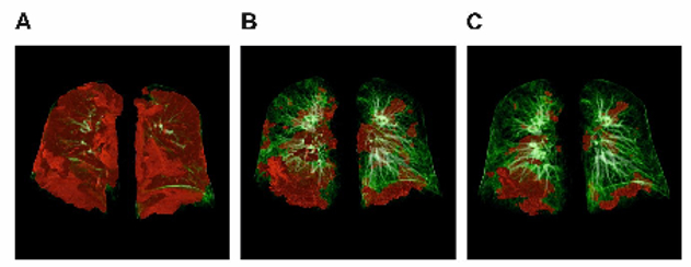
Fig. 1: Sequential CT scans of a 56-year old male COVID-19 patient during acute disease and follow-up. Pulmonary 3D modelling assessed with CT during (A) acute COVID-19, at (B) 60 days follow-up and at (C) 100 days follow-up is shown. Pulmonary opacities, mainly reflecting ground-glass opacities and/or consolidation, were quantified with Syngo.via CT Pneumonia Analysis software. Areas with increased opacity are marked with red colour, whereas normal lung areas are indicated in green.
c) Low dose imaging
- ALADA (as low as diagnostically acceptable)- dose management of CT-guided interventions of the lumbar spine
- Prediction of ALADA Doses for Implant Site Analysis Using Slope of Hounsfield Units- external collaboration with King Saud University
- Development of an ALADA reference quality approach in craniofacial trauma CT
- Clinical verification of an ALADA protocol in craniofacial trauma
EXPERIMENTAL RADIOLOGY
a) Image Processing
Wolfgang Recheis
Image processing and analysis, including rapid prototyping based on radiological data, represent core interests and tasks, including multidimensional visualisation, the quantification of disease patterns based on texture analysis, shape analysis and others. Moreover, our new micro-CT core facility allows depiction of structures on μm scale in all three spatial dimensions.
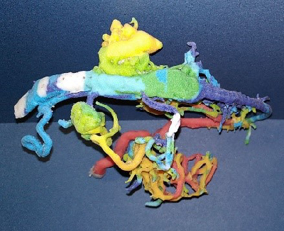
Fig. 2: Full-colour 3D printout of an arteriovenous malformation (AVM) on the basis of 2D angiography, mapped on 3D CT
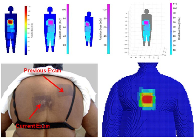
Fig. 3: Screenshots of our in-house developed dose management system “SumDose”: a real-time dose-monitoring system, work-in-progress of the dose management group
b) Small Animal MRI
Christian Kremser
In addition to clinically oriented projects, the Department of Radiology conducts experimental MR examinations on various animal models and specimens. Over the years, coil setup and imaging sequences have been optimised accordingly. Study topics include: mouse spinal cord imaging after shock wave treatment to promote spinal cord repair; volumetry of mouse myocardium to quantify fibrotic changes after constriction of the aorta and pulmonary artery; volumetry of the mouse brain to quantify fixation shrinkage; pulse wave velocity imaging in rabbits to evaluate aortic plaques; volumetric quantification of subcutaneous and visceral fat in mice on different diets; detection of intervertebral disc herniation in knockout mice.
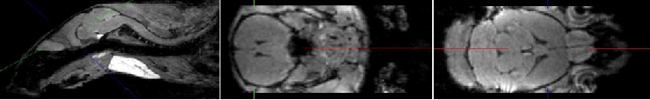
Fig. 4: Examples of mouse brain MR images obtained in 3T with an isotropic resolution of 200µm.
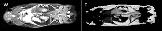
Fig. 5: Examples of mouse whole-body MR images obtained in 3T for quantification of subcutaneous and visceral fat.
MedCorpInn- Retrospective Intersectional Corpuslinguistic Analysis of Radiological and Medical Reports of Medical University of Innsbruck
Claudia Posch and Karoline Irschara (University of Innsbruck), Leonhard Gruber and Stephanie Mangesius (Medical University of Innsbruck)
This project is compiling a large linguistic corpus (digitalised data collection) of radiology reports and medical reports, in order to conduct corpus and discourse linguistic as well as gender medicine research. After standardisation, automated data processing and anonymisation, the radiology corpus contains over 5 million anonymised radiology reports, which can be queried using corpus linguistic and NLP tools. With respect to linguistic patterns and differences, this unique data set can be used e.g. to study whether differences in language use are connected with social and demographic factors.
Abdominal MR Imaging
Michaela Plaikner, Benjamin Henninger, Christian Kremser
Morphological and functional MRI in all-organ systems development of novel MRI applications and MR sequences. Examples of research projects: fat, iron or combined disease; influence of iron on the evaluation of liver fat.
a) MRI for the evaluation of diffuse liver disease:
evaluation of different MRI methods (relaxometry, chemical shift imaging, multi-echo approach, Dixon screening), in order to detect diffuse liver disease (fat, iron or combined disease); influence of iron on the evaluation of liver fat.
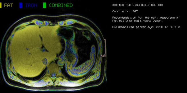
Fig. 6: Left: 43year old patient with fatty liver.
b) MR-Elastography (MRE)
MRE is increasingly used in hepatic MRI to detect and classify fibrosis in the early stages before morphological changes have occurred. In our department, MRE is already integrated into the routine hepatic MRI protocol and used for various research projects.
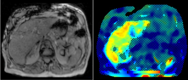
Fig. 7: Example of hepatic MRE. The colour image represents the stiffness map obtained. In this case, the mean hepatic stiffness was 4.83kPa, which indicates grade F3 fibrosis.
Cardiovascular MR Imaging
Agnes Mayr, Christian Kremser, in cooperation with the Department of Cardiology
a) STEMI CMR: CMR Parameters of Myocardial Tissue Damage in ST-Elevation Myocardial Infarction (STEMI).
Since 2005, almost 900 patients have been examined under a comprehensive cardiac MRI (CMR) protocol within the first week as well as 4 months, 12 months and 10 years after acute STEMI. In more than 60 internal original papers, CMR myocardial infarction severity markers were assessed and the effects of CMR on optimised risk assessment shortly after STEMI were evaluated.
b) TAVI CMR: CMR to Guide Transcatheter Aortic Valve Implantation (TAVI).
This ongoing randomised study investigates the non-inferiority of TAVI CMR to TAVI CT for the first time, with regard to efficacy and safety end-points in the guidance for TAVI evaluation.
c) 4D Phase Contrast Flow Measurements
- Patients with different grades of aortic valve stenosis: comparison of 4D flow-assessed stenosis severity with 3D echocardiography and invasive measurements.
- Patients with cryptogenic stroke: recording turbulent kinetic energy, changes in flow patterns and regional wall stresses as well as occurrence of a vortex-shaped flow along the thoracic aortic wall, in order to optimise the elucidation of potential cardioembolic sources.
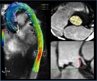
Fig. 8: 4D Phase contrast flow measurement of the thoracic aorta (a). Imaging example of non-contrast 3D “whole-heart” CMR for (b) aortic annular measurements in the hinge-point plane and (c) left main artery ostial height measurement for prosthesis sizing.
Imaging and Targeted Biopsy of the Genitourinary (GU) Tract
Friedrich Aigner
- Multiparametric ultrasound of the scrotum
- Multiparametric ultrasound of the prostate
- Multiparametric MRI of the prostate
- Fusion imaging of the GU-tract
- Fusion targeted biopsy of the prostate
The simultaneous application of structural and functional imaging techniques is described as multiparametric (MP) (Fig. 9). Studies have shown that the MP approach results in greater diagnostic accuracy (Fig. 10).
The use of fusion imaging in uroradiology improves ultrasound lesion-detection rates, shows more reliable size controls at different time points, is an alternative to in-bore biopsies (Fig. 11) and can be used for focal therapy.
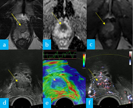
Fig. 9: Multiparametric MRI (a – c) and multiparametric transrectal ultrasound (d – f) of the prostate in the same patient show a histologically confirmed prostate carcinoma (arrows) as an area of low signal intensity / low echogenicity in T2-weighted MRI (a) and B-mode scan (d), with increased cell density in diffusion imaging (b, ADC-MAP) and ultrasound elastography (e, colour-coded blue), and as an area of hyperperfusion in contrast-enhanced MRI (c) and Doppler ultrasound (f).
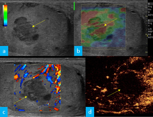
Fig. 10: Patient with suspected testicular cancer: the multiparametric ultrasound of the testicle shows (a, arrow) a hypoechoic suspected cancerous lesion in B-scan, which is (b, arrow) soft in elastography (colour coded in green and red) and (c, d) avascular in (c) Doppler and (d) contrast medium ultrasound. In the light of these mpUS features, the new working diagnosis was changed to “cancer-simulating ischaemic orchitis”.
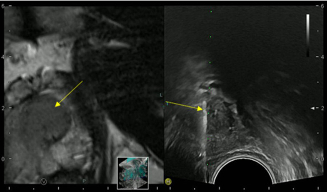
Fig. 11: US/MRI fusion-targeted biopsy of the prostate: in the left image, T2-weighted MRI shows the prostate carcinoma as an area of low signal intensity that also infiltrates the urethra (arrow); the right image shows the real-time ultrasound in B-mode on the split ultrasound monitor as well as the correct needle position (arrow) whilst performing the ultrasound-guided biopsy of the suspicious MRI lesion. Histology revealed carcinoma of the prostate with a Gleason score of 8 (4 + 4).
Musculoskeletal Imaging
Andrea S. Klauser et al., Multicentre collaboration for indication in MSK imaging and interventions
- Sonography of carpal tunnel: definition of cut-off values
- Sonoelastography of epicondylitis, plantar fasciitis and Achilles tendon: accuracy in comparison with histology
- Sonographically guided (SG) injections in CTS: sonoelastographic appearance
- SG peripheral nerve injection: sonomorphology
- SG nerve entrapment syndromes in comparison with surgical outcome
- SG injection in sacroiliac joints of children: to prove feasibility
- Dual-energy CT (DECT) in gout: comparison with US, findings in extra-articular regions and cardio in gout and subgroup patients
- X-ray in comparison with DECT in gout patients
- X-ray in comparison with US in erosion assessment
- MRI in comparison with US in tendon overuse assessment
- MRI whole-spine imaging in inflammatory disease: therapeutic follow-up assessment
- MRI sacroiliitis: therapeutic follow-up assessment
- MR tractography (DTI, ADI) in median nerves of healthy volunteers and CTS patients: comparison with sonography.
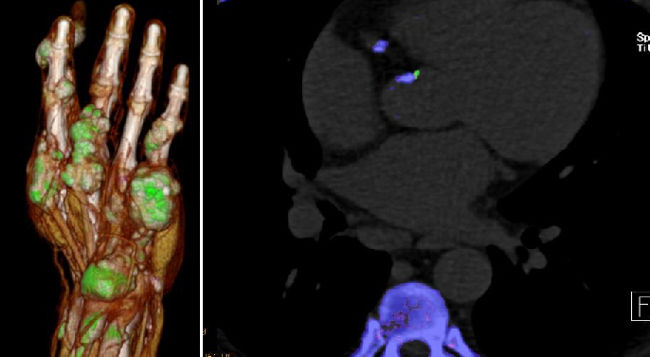
Fig. 12: Left: Gout deposits (in green) detected by DECT; Right: Gout deposits (green) in coronary arteries detected by DECT
Non-Invasive Cardiac/Cardiovascular CT Imaging
Gudrun Feuchtner
a) Coronary heart disease: atherosclerosis, high-risk plaque marker, risk estimation, algorithms, non-invasive FFR
b) Structural heart disease CT for planning TAVR, TMVR and catheter ablation, including 3D printing, advanced 3D visualisation, congenital heart disease (Fig. 13).
CT: 10 Multicentre trials
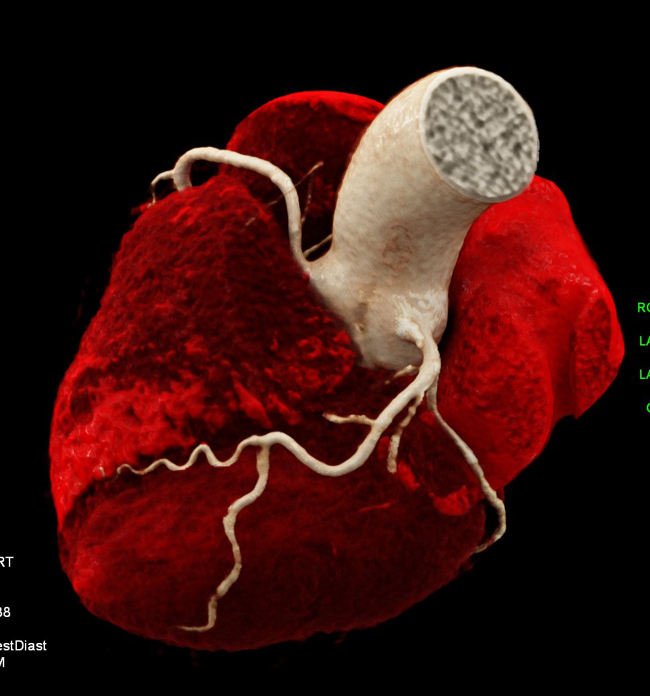
Fig. 13: 3D Print of coronary arteries after image post-processing.
c) Atherosclerotic Burden and its Relevance to Different Diseases and Treatment Strategies.
Bernhard Glodny, Johannes Petersen
Cardiovascular and cerebrovascular sequelae of atherosclerotic disease are among the leading causes of morbidity and mortality in humans. Atherosclerosis can be detected using different modalities of diagnostic imaging. Moreover, treatments are planned by means of clinical imaging, and cardiovascular risk profiles can be compiled individually. CT can be used to quantify the “atherosclerotic burden” of all vascular territories in an objective and reproducible manner.
Relationships between oral health and health in general have been suspected for many years and could also be proven by imaging studies, with evidence of an association between inflammatory changes in the periodontium and increased arteriosclerosis load, thus supporting the concept of arteriosclerosis as an inflammatory disease.
Ultrasound
Hannes Gruber, Alexander Loizides
- Peripheral nerve sonography
- SG interventions in the peripheral nervous system
- SG carpal tunnel release
- Sonographic evaluation of soft tissue masses (incl. MSK contrast-enhanced sonography (CEUS))
- Sonography of the musculoskeletal system
- Sonographic assessment of functional musculoskeletal disorders
- SG pain therapy
- Angiologic assessment and SG interventions in the vascular system
The surgical ultrasound section is a leader in the development of ultrasound techniques for the evaluation of peripheral nerves, ultrasound-guided nerve root infiltration and pain therapy. One of the most recent publications illustrates our work.
The axillary nerve (AN) is frequently injured during shoulder trauma and imaging is required to define the site and extent of nerve injury. However, the AN has a rather complex course through several soft tissue compartments of the shoulder and axilla. Detection and sonographic assessment therefore require thorough knowledge of the local topography.
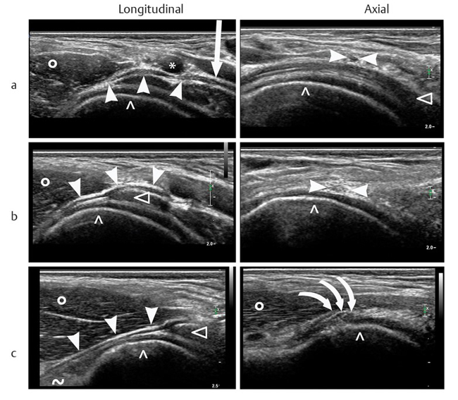
Fig. 14: The 2 US scans a (longitudinal and axial) show the sonographic situation at the “departure” of the AN from the plexus, the 2 US scans b (longitudinal and axial) show the sonographic situation at the “middle segment”, and the 2 US scans c (longitudinal and axial) show the sonographic situation at the “distal segment”. The white arrowheads indicate the AN, the white arrow indicates the posterior fascicle of the plexus, (*) indicates the axillary artery, (^) indicates the cortex of the humeral head, (Δ) indicates the subscapular muscle, the rings (0) indicate the teres minor muscle, (~) indicates the long head of the triceps muscle, and the 3 bowed white arrows indicate the 3 terminal branches of the AN forming within the axillary gap.
Our investigation aims to define reliable, anatomical landmarks for AN sonography in volunteers and later to validate the proposed sonographic examination protocol in patients. With strict adherence to the proposed examination algorithm, sonography of the AN was feasible in all volunteers and patients. The findings correlated nicely with the gold standard of “surgical exploration” in respect of the severity and topography of neural impairment.
Based on our study results, we propose our algorithm for AN sonography as the first-line imaging tool for the assessment of axillary nerve trauma (PIC).
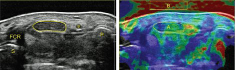
Fig. 15: Carpal tunnel syndrome: diagnosis by means of median nerve elasticity—improved diagnostic accuracy of US with sonoelastography (Miyamoto et al., 2014).
Interventional Oncology
Reto Bale
- Image-guided tumour ablation
- Stereotaxis
- Robotics
- Targeting
- Interventional oncology
Stereotactic ablation of liver tumours:
Radiofrequency ablation (RFA) allows local curative tumour treatment by inducing coagulation necrosis with a high-frequency alternating current. The major limiting factor of conventional US- and CT- guided percutaneous single probe ablation is the tumour size. Navigation systems allow for 3D-planning of multiple overlapping ablation zones on the CT datasets and precise transformation into the real patient. We developed the worldwide first aiming device for frameless stereotactic punctures and performed the first in man stereotactic radiofrequency ablation (SRFA) of a liver tumor in 2001. Meanwhile > 1000 patients with > 3000 liver tumors, most of them being inoperable, have successfully been treated at our department.
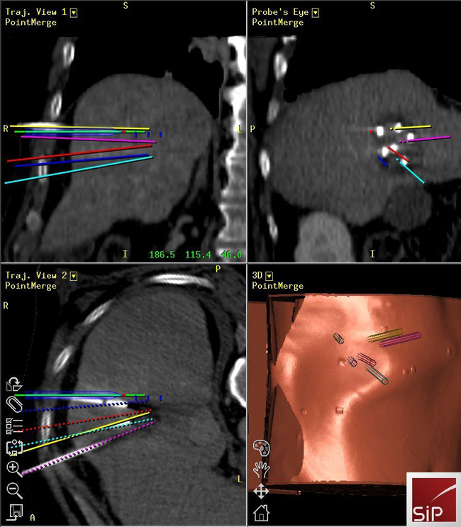
Fig. 16: SRFA of a liver tumor with verification of precise needle placement by image fusion (screenshot of 3d navigation system)
Imaging and Targeted Biopsy of Breast Cancer
Martin Daniaux
Dual-energy (DE) contrast-enhanced mammography is one of the latest developments in breast care. Imaging with contrast agents in breast cancer has been described in MRI and CT. However, high costs, limited availability and high radiation doses have led to the development of contrast-enhanced spectral mammography (CESM).
The research team focusing on the diagnosis of breast cancer has a vast wealth of experience with all the imaging modalities currently used for evaluation of the breast, including fine-needle aspiration and/or percutaneous biopsies using stereotaxis or ultrasound guidance. Our unit serves as the largest screening and assessment centre of the national breast-screening programme in Tyrol. Approximately 10,000 mammograms and breast ultrasound studies are performed each year.
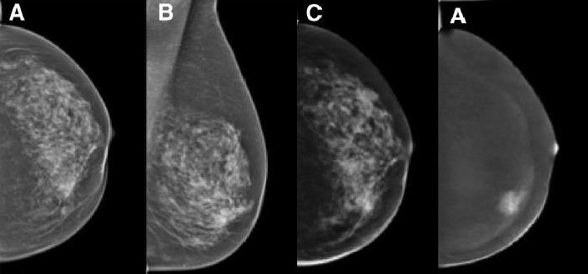
Fig. 17: woman with an invasive ductal carcinoma, showing inhomogeneous breast tissue at 9 o’clock, as demonstrated by conventional mammography (a, b). Low-energy CESM images (c) are equivalent to conventional mammography, whereas spectral imaging shows a contrast-enhancing mass.
Selected Publications
CT Imaging
- Riechelmann H, Widmann G, Kofler B, Arminger R, Url C, Giotakis AI.: Nasal Floor Asymmetry Is Associated With Nasal Obstruction. J Oral Maxillofac Surg. 2020 May 14:S0278-2391(20)30464-X.
- Dejaco D, Steinbichler T, Schartinger VH, Fischer N, Anegg M, Dudas J, Posch A, Widmann G, Riechelmann H.: Specific growth rates calculated from CTs in patients with head and neck squamous cell carcinoma: a retrospective study performed in Austria. BMJ Open. 2019 Jul;98(7):500-502.
- Sonnweber T, Boehm A, Sahanic S, Pizzini A, Aichner M, Sonnweber B, Kurz K, Koppelstätter S, Haschka D, Petzer V, Hilbe R, Theurl M, Lehner D, Nairz M, Puchner B, Luger A, Schwabl C, Bellmann-Weiler R, Wöll E, Widmann G, Tancevski I, Judith-Löffler-Ragg, Weiss G.: Persisting alterations of iron homeostasis in COVID-19 are associated with non-resolving lung pathologies and poor patients' performance: a prospective observational cohort study. Respir Res. 2020 Oct 21;21(1):276.
Experimental Radiology
- Gollmann-Tepeköylü C, Nägele F, Graber M, Pölzl L, Lobenwein D, Hirsch J, An A, Irschick R, Röhrs B, Kremser C, Hackl H, Huber R, Venezia S, Hercher D, Fritsch H, Bonaros N, Stefanova N, Tancevski I, Meyer D, Grimm M, Holfeld J.: Shock waves promote spinal cord repair via TLR3. JCI Insight. 2020; 5(15): e134552.
- García-Martínez, D., Bastir, M., Villa, C. Daniel García-Martínez, Markus Bastir, Chiara Villa, Francisco García-Río, Isabel Torres-Sánchez, Wolfgang Recheis, Alon Barash, Roman Hossein Khonsari, Paul O’Higgins, Marc R. Meyer & Yann Heuzé.: Late subadult ontogeny and adult aging of the human thorax reveals divergent growth trajectories between sexes. Sci Rep 2020, Jul 1;10(1):10737.
- Verius M, Frank F, Gizewski E, Broessner G.: Magnetic Resonance Spectroscopy Thermometry at 3 Tesla: Importance of Calibration Measurements. Ther Hypothermia Temp Manag. 2019;9(2):146-155.
Abdominal MR-Imaging
- Henninger B, Alustiza J, Garbowski M, Gandon Y.: Practical guide to quantification of hepatic iron with MRI. Eur Radiol. 2020 Jan;30(1):383-393. doi: 10.1007/s00330-019-06380-9.
- Henninger B, Plaikner M, Zoller H, Viveiros A, Kannengiesser S, Jaschke W, Kremser C.: Performance of different Dixon-based methods for MR liver iron assessment in comparison to a biopsy-validated R2* relaxometry method. Eur Radiol. 2020 Sep 23. doi: 10.1007/s00330-020-07291-w. Online ahead of print.
- Plaikner M, Kremser C, Zoller H, Steurer M, Glodny B, Jaschke W, Henninger B.: Does gadoxetate disodium affect MRE measurements in the delayed hepatobiliary phase? Eur Radiol. 2019 Feb;29(2):829-837.
Cardiovascular MR-Imaging
- Mayr A, Pamminger M, Reindl M, Greulich S, Reinstadler SJ, Tiller C, Holzknecht M, Nalbach T, Plappert D, Kranewitter C, Klug G, Metzler B.: Mitral annular plane systolic excursion by cardiac MR is an easy tool for optimized prognosis assessment in ST-elevation myocardial infarction. Eur Radiol. 2020 Jan;30(1):620-629
- Pamminger M, Klug G, Kranewitter C, Reindl M, Reinstadler SJ, Henninger B, Tiller C, Holzknecht M, Kremser C, Bauer A, Jaschke W, Metzler B, Mayr A.: Non-contrast MRI protocol for TAVI guidance: quiescent-interval single-shot angiography in comparison with contrast-enhanced CT. Eur Radiol. 2020 Sept; 30(9): 4847-4856
- Mayr A, Klug G, Reinstadler SJ, Feistritzer HJ, Reindl M, Kremser C, Kranewitter C, Bonaros N, Friedrich G, Feuchtner G, Metzler B.: Is MRI equivalent to CT in the guidance of TAVR? A pilot study. Eur Radiol. 2018 Nov;28(11):4625-4634
Imaging and Targeted Biopsy of the Genitourinary (GU) Tract
- Auer T, Heidegger I, DE Zordo T, Junker D, Jaschke W, Steinkohl F, Aigner F.: Fusion Imaging of Contrast-enhanced Ultrasound With CT or MRI for Kidney Lesions. In Vivo. 2019 Jan-Feb;33(1):203-208. doi: 10.21873/invivo.11460.
- Steinkohl F, Luger AK, Pichler R, Bektic J, Rehder P, Lebovici A, Aigner F.: Visibility of MRI prostate lesions on B-mode transrectal ultrasound. Med Ultrason. 2018 Dec 8;20(4):441-445. doi: 10.11152/mu-1602.
- Auer T, De Zordo T, Dejaco C, Gruber L, Pichler R, Jaschke W, Dogra VS, Aigner F.: Value of Multiparametric US in the Assessment of Intratesticular Lesions. 2017 Nov;285(2):640-649. doi: 10.1148/radiol.2017161373. Epub 2017 Jun 16.
Musculoskeletal Imaging
- Klauser AS, Halpern EJ, Strobl S, Gruber J, Feuchtner G, Bellmann-Weiler R, Weiss G, Stofferin H, Jaschke W.: Dual-Energy Computed Tomography Detection of Cardiovascular Monosodium Urate Deposits in Patients With Gout. JAMA Cardiol. 2019 Oct 1;4(10):1019-1028.
- Strobl S, Kremser C, Taljanovic M, Gruber J, Stofferin H, Bellmann-Weiler R, Klauser AS.: Impact of Dual-Energy CT Postprocessing Protocol for the Detection of Gouty Arthritis and Quantification of Tophi in Patients Presenting With Podagra: Comparison With Ultrasound. AJR Am J Roentgenol. 2019 Dec;213(6):1315-1323.
- Sconfienza LM, Adriaensen M, Albano D, Allen G, Aparisi Gómez MP, Bazzocchi A, Beggs I, Bignotti B, Chianca V, Corazza A, Dalili D, De Dea M, Del Cura JL, Di Pietto F, Drakonaki E, Facal de Castro F, Filippiadis D, Gielen J, Gitto S, Gupta H, Klauser AS, Lalam R, Martin S, Martinoli C, Mauri G, McCarthy C, McNally E, Melaki K, Messina C, Mirón Mombiela R, Neubauer B, Olchowy C, Orlandi D, Plagou A, Prada Gonzalez R, Rutkauskas S, Snoj Z, Tagliafico AS, Talaska A, Vasilevska-Nikodinovska V, Vucetic J, Wilson D, Zaottini F, Zappia M, Obradov M.: Clinical indications for image-guided interventional procedures in the musculoskeletal system: a Delphi-based consensus paper from the European Society of Musculoskeletal Radiology (ESSR)-part I, shoulder. Eur Radiol. 2020 Feb;30(2):903-913.
Non-invasive Cardiac/Cardiovascular CT-Imaging
- Feuchtner GM, Barbieri F, Luger A, Skalla E, Kountchev J, Widmann G, Plank F.: Myocardial injury in COVID-19: The role of coronary computed tomography angiography (CTA). J Cardiovasc Comput Tomogr. 2020 Jul 17:S1934-5925(20)30391-9. Epub ahead of print.
- Feuchtner G, Langer C, Barbieri F, Beyer C, Dichtl W, Friedrich G, Schgoer W, Widmann G, Plank F.: The effect of omega-3 fatty acids on coronary atherosclerosis quantified by coronary computed tomography angiography. Clin Nutr. 2020 Jul 22:S0261-5614(20)30381-2.
- van Rosendael AR, Narula J, Lin FY, van den Hoogen IJ, Gianni U, Al Hussein Alawamlh O, Dunham PC, Peña JM, Lee SE, Andreini D, Cademartiri F, Chinnaiyan K, Chow BJW, Conte E, Cury RC, Feuchtner G, Hadamitzky M, Kim YJ, Leipsic J, Maffei E, Marques H, de Araújo Gonçalves P, Plank F, Pontone G, Raff GL, Villines TC, Weirich HG, Al'Aref SJ, Baskaran L, Cho I, Danad I, Han D, Heo R, Lee JH, Rivzi A, Stuijfzand WJ, Gransar H, Lu Y, Sung JM, Park HB, Samady H, Stone PH, Virmani R, Budoff MJ, Berman DS, Chang HJ, Bax JJ, Min JK, Shaw LJ.: Association of High-Density Calcified 1K Plaque With Risk of Acute Coronary Syndrome. JAMA Cardiol. 2020 Mar 1;5(3):282-290
Ultrasound
- Gruber H, Jaschke W, Loizides A.: Ultrasound Guidance for Pleural-Catheter Placement. N Engl J Med. 2018 Jul 26;379(4):401.
- Gruber L, Jimenez-Franco LD, Decristoforo C, Uprimny C, Glatting G, Hohenberger P, Schoenberg SO, Reindl W, Orlandi F, Mariani M, Jaschke W, Virgolini IJ.: MITIGATE-NeoBOMB1, a Phase I/IIa Study to Evaluate Safety, Pharmacokinetics and Preliminary Imaging of 68 Ga-NeoBOMB1, a Gastrin- releasing Peptide Receptor Antagonist, in GIST Patients. J Nucl Med. 2020. Epub ahead of print.
Interventional Oncology
- Bale R, Schullian P, Eberle G, Putzer D, Zoller H, Schneeberger S, Manzl C, Moser P, Oberhuber G.: Stereotactic Radiofrequency Ablation of Hepatocellular Carcinoma: a Histopathological Study in Explanted Livers. Hepatology. 2019 Sep;70(3):840-850.
- Schullian P, Johnston EW, Putzer D, Laimer G, Waroschitz G, Braunwarth E, Amann A, Maglione M, Bale R.: Stereotactic radiofrequency ablation (SRFA) for recurrent colorectal liver metastases after hepatic resection. Eur J Surg Oncol. 2020 Oct 1:S0748-7983(20)30806-4.
- Laimer G, Schullian P, Jaschke N, Putzer D, Eberle G, Alzaga A, Odisio B, Bale R.: Minimal ablative margin (MAM) assessment with image fusion: an independent predictor for local tumor progression in hepatocellular carcinoma after stereotactic radiofrequency ablation. Eur Radiol. 2020 May;30(5):2463-2472
Imaging and Targeted Biopsy of Breast Cancer
- Geiger-Gritsch S, Daniaux M, Buchberger W, Knapp R, Oberaigner W.: Performance of 4 years of population-based mammography screening for breast cancer combined with ultrasound in Tyrol / Austria. Wien Klin Wochenschr. 2018 Feb;130(3-4):92-99.
- van Dam PA, Tomatis M, Marotti L, Heil J, Mansel RE, Rosselli Del Turco M, van Dam PJ, Casella D, Bassani LG, Danei M, Denk A, Egle D, Emons G, Friedrichs K, Harbeck N, Kiechle M, Kimmig R, Koehler U, Kuemmel S, Maass N, Mayr C, Prové A, Rageth C, Regolo L, Lorenz-Salehi F, Sarlos D, Singer C, Sohn C, Staelens G, Tinterri C, Audisio R, Ponti A; eusomaDB Working Group.: Time trends (2006-2015) of quality indicators in EUSOMA-certified breast centres. Eur J Cancer. 2017 Nov;85:15-22.
- Oberguggenberger A, Martini C, Huber N, Fallowfield L, Hubalek M, Daniaux M, Sperner-Unterweger B, Holzner B, Sztankay M, Gamper E, Meraner V.: Self-reported sexual health: Breast cancer survivors compared to women from the general population - an observational study. BMC Cancer. 2017 Aug 30;17(1):599.
Selection of Funding
Selection of Competitive Acquired Funding
- MITIGATE #602306: Closed-loop Molecular Environment for Minimally Invasive Treatment of Patients with metastatic Gastrointestinal Stromal Tumours; EU FP7; Univ.-Prof. Dr. Werner Jaschke
- EUCLID: European Study on Clinical Diagnostic Reference Levels for X-ray Medical Imaging; Europ. Commission: Contracnr. EC Tender ENER/2017/NUCL/SI2.759174; Univ.-Prof. Dr. Elke R. Gizewski, em.o.Univ.-Prof. Dr. Werner Jaschke, Dr. Michael Verius
- VASCage-C: Imaging Biomarkers For Vascular Diseases And Vascular Aging; COMET K1 Zentren 4.Call: 1. FP Partner (FFG), Projektnummer: 6401616; Univ.-Prof. Dr. Elke R. Gizewski, Assoz.-Prof. PD Dr. Bernhard Glodny, PD Dr. Wolfgang Recheis, Assoz.-Prof. PD Dr. Astrid E. Grams, Dr. Ruth Steiger, Dr. Sergiy Pereverzyev Jr.
- Subproject Vascage: Post-Stroke Osteopathie; Degenhart Gerald MSc BSc
- KM-induziertes Nierenversagen bei chron. Niereninsuffizienz im Schweinemodell: „CIN- Pig“; MFF Tirol #265; Dr. Anna-Katharina Luger, Assoz.-Prof. PD Dr. Bernhard Glodny
- Basic evaluation of a contrast-free pre-TAVI imaging option: first comparison of a new native MRI protocol with standard-ized contrast-enhanced CT; GHKF Gesellschaft zur Förderung der Herz-Kreislaufforschung in Tirol; Assoz.-Prof. PD Dr. Agnes Mayr
- Long term evolution of infarct scar after reperfused ST-segment elevation myocardial infarction: a 10 year Follow-Up Cardiac Magnetic Resonance Study; ÖKG Österreichische Kardiologische Gesellschaft; Assoz.-Prof. PD Dr. Agnes Mayr
- DISCHARGE Trial : Diagnostic Imaging Strategies: Invasive versus coronary computed tomography angiography for stable chest pain patients with intermediate risk of coronary artery disease: a multicentric randomized pragmatic trial.; 7th Framework Programme (Grant No. 603266) of the European Union.; Ao. Univ.-Prof. Dr. Gudrun Feuchtner
- MedCorpInn - Retrospective Intersectional Corpuslinguistic Analysis of Radiology and Medical Reports of Medical University of Innsbruck; Austrian Academy of Sciences, go!digital 2.0; Mag. Dr. Claudia Posch (University of Innsbruck, Institute for Linguistics), Karoline Irschara, MA (University of Innsbruck, Institute for Linguistics), Dr. med. univ. Leonhard Gruber, PhD (Universitätsklinik für Radiologie, Medizinische Universität Innsbruck), Dr.in med. univ. Stephanie Mangesius, PhD (Universitätsklinik für Neuroradiologie, Medizinische Universität Innsbruck)
Collaborations
- Siemens Healthcare Erlangen
- Interventional Systems, Kitzbühel
- Medtronic, Boulder, USA
- Leopold Franzens Universität Innsbruck: Institut für Geologie : Fachbereich Sedimentologie: Univ.-Prof. Dr. Michael Strasser; Institut für Mineralogie und Petrographie: Ao.Univ.-Prof. Dr. Peter Tropper; Institut für Archäologien : Fachbereich Ur-und Frühgeschichte sowie Mittelalter- und Neuzeitarchäologie: Univ.-Prof. Dr. Harald Stadler; Fachbereich Klassische und Provinzialrömische Archäologie: Mag. Dr. Ulrike Töchterle, Mag. Dr. Martin Auer; Institut für Sprachwissenschaft: Ass.Prof. Mag. Dr. Claudia Posch, Karoline Irschara, MA; Applied Mathematics: Univ.-Prof. Markus Haltmeier
- Eidgenössische Technische Hochschule Zürich: Institut für Biomechanik: Univ. Prof. Dr. Ralph Müller
- Universitätsklinikum Ulm: Institut für Unfallchirurgische Forschung und Biomechanik: Univ. Prof. Dr. Anita Ignatius
- Johannes Keppler Universität: Institut für Physiologie & Pathophysiologie: Univ. Prof. Dr. Jakob Völkl
- Landesmuseum Ferdinandeum: Mag.phil. Wolfgang Sölder
- Kelten Museum Hallein: Margarethe Kirchmayr, MA
- National Taiwan University: Jyh-Jaan Huang PHD (momentan Forschungsaufenthalt an der LFU Innsbruck)
- ASTC Axel Schnaller Technologie Consult
- Managementcenter Innsbruck: Mechatronik- Medizintechnik: Dr. techn. Franz- Josef Falkner
- Cardiac gout project with DECT: multicenter collaboration with Rheumatology Department at Vancouver General Hospital (VGH) and Massachusetts General Hospital (MGH)
- MSK ultrasound collaborations with Thomas Jefferson University, PA and Department of Medical Imaging, The University of Arizona Health Network
- MSK Imaging collaborations with Diagnostic and Interventional Radiology, Istituto Ortopedico Galeazzi, Italy
- Novartis Institutes for Biomedical research, Biomarker Development- Clinical Imaging
- Basel, Switzerland: Tendon stiffness as a biomarker to quantify tendon healing after Achilles tendon rupture (2016). Projektnummer (Powertrials 1302): Director Didier Laurent, PhD
- King Saud University: Prof. Dr. Asma’a al Ekrish Division of Oral and Maxillofacial Radiology, Department of Oral Medicine and Diagnostic Sciences, College of Dentistry, King Saud University, Riyadh, Saudi Arabia
Devices & Services
- Corefacility Micro- CT
- Administrative Director: Gerald Degenhart, BSc, MSc
- Scientific Director: Priv.-Doz. MMag. Dr. Johannes D. Pallua, PhD
 Univ.-Prof.in Dr.in med. Elke R. Gizewski, MHBA
Director (interim)
Contact:
Anichstraße 35
6020 Innsbruck
Austria
E-mail: radiologie@i-med.ac.at
Phone: +43 512 504 22798
Fax: +43 512 504 22758
https://radiologie.tirol-kliniken.at/page.cfm?vpath=radiologie/forschung/micro-ct